Flow Cytometry Leukemia Lymphoma Panel
Flow cytometry immunophenotyping may be useful in helping to diagnose classify treat and determine prognosis of these blood cell cancers. Flow cytometry is an indispensable tool for diagnosis and monitoring of leukemia and lymphoma.
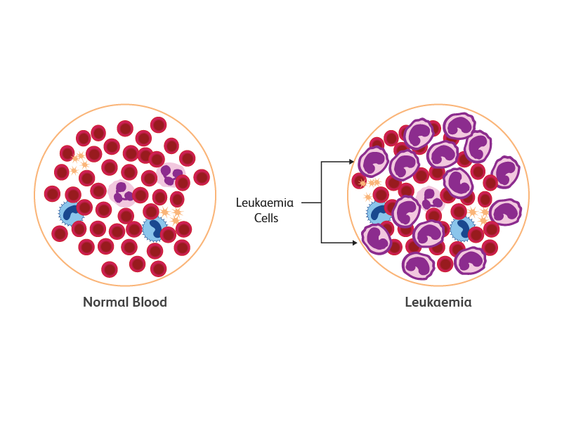
What Is Leukaemia Types Of Leukaemia Leukaemia Causes
LEUKEMIALYMPHOMA PANEL General Information Lab Order Codes.

Flow cytometry leukemia lymphoma panel. Immunophenotyping based on CD45 gating. Flow cytometry plays an important role in the diagnosis monitoring and treatment of haematological malignancies. Most consensus leukemia lymphoma antibody panels consist of lists of markers based on expert opinions but they have not been validated.
The resulting CD expression or immunophenotype is used by the hematopathologist to aid in the identification and diagnosis of various malignancies as well as to follow up after treatment. In addition to reflexing flow cytometric panels AML FISH testing for PML-RARA translocation t1517 may be added by the Mayo Clinic pathologist to exclude acute promyelocytic leukemia if there is morphologic suspicion or if blasts and promyelocytes are CD34-negative and HLA-DR-negative. Flow cytometry now used routinely to aid in the classification of leukemias is increasingly being evaluated as a rapid technique for determination of surface antigens on the cells teased from lymph nodes and other masses with suspected lymphoma.
In addition to reflexing flow cytometric panels fluorescence in situ hybridization FISH or molecular testing may be recommended by the Mayo pathologist to facilitate diagnosis. Flow cytometric leukemia and lymphoma analysis may aid in identifying the tumor lineage for diagnostic and prognostic purposes. For solid tissue specimens order LLPT LeukemiaLymphoma Immunophenotyping Flow Cytometry Tissue.
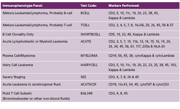
Flow Cytometry Test Description Markers are CD2 CD3 CD4 CD5 CD7 CD8 CD10 CD11b CD11c CD13 CD14 CD15 CD16 CD19 CD20 CD23 CD33 CD34 CD38 CD41 CD45 CD56 CD64 CD71 CD117 CD138 CD235a FMC-7 HLA-DR kappa and lambda. Testing begins with decisions about which screen test panels to use for individual samples as they are received by the laboratory. After review of the clinical history and morphology a panel of markers is selected for each case by a board-certified hematopathologist.
For bone marrow specimens being evaluated for possible involvement by a myelodysplastic syndrome MDS or a myelodysplasticmyeloproliferative neoplasm MDSMPN including chronic myelomonocytic leukemia CMML order MYEFL Myelodysplastic Syndrome by Flow Cytometry Bone Marrow. They will contact the referring physician or pathologist to confirm the addition of these tests. While application of flow cytometry in this field may be complex and require a lot of experience it is based on rather simple principles.
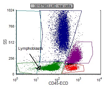
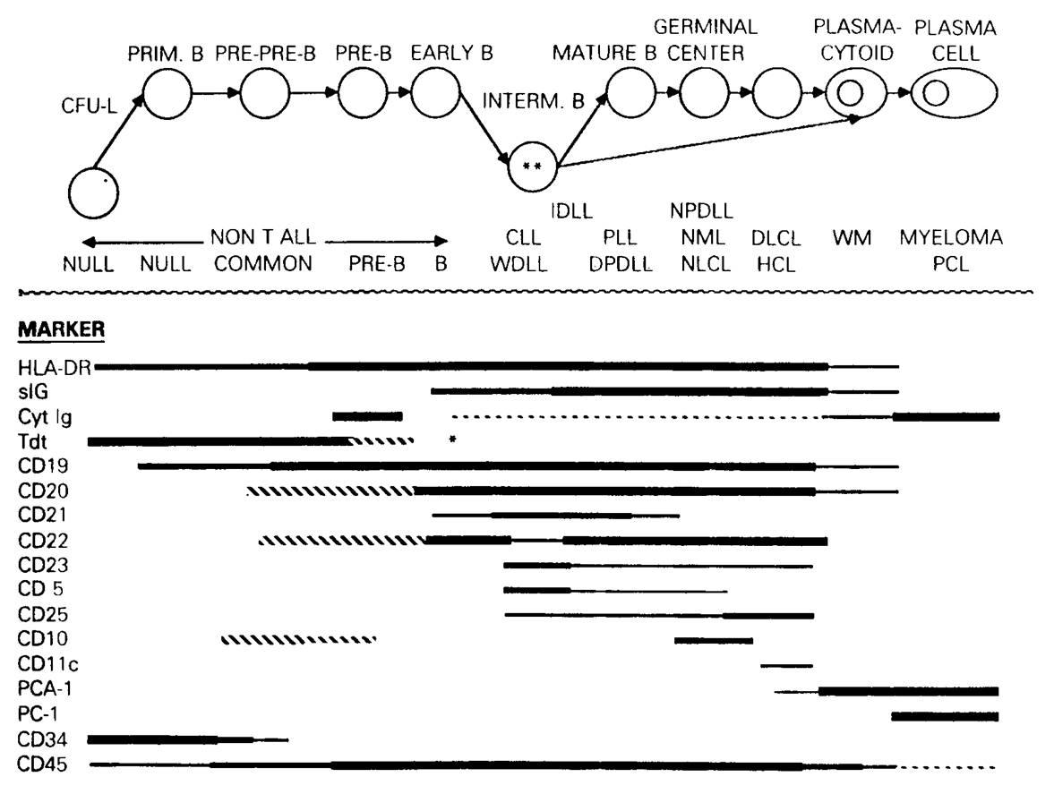
LeukemiaLymphoma B-ALL with Hematogone Hyperplasia 2019 MFMER slide-14 B-Lymphoblasts and Hematogones Hematogones show a reproducible maturation pattern B-lymphoblasts essentially always demonstrate immunophenotype aberrancies Beware of hematogone hyperplasia in the setting of B-ALL Flow cytometry and morphology usually provide concordant blast percentages Hemodilution. Leukemias and lymphomas are caused by an abnormal white blood cell that begins to divide uncontrollably making numerous copies of itself clones. Nine-color and 10-color flow cytometry offers the possibility for increased accuracy in population identification the ability to obtain detailed information from paucicellular specimens improved laboratory efficiency and the means to consistently detect abnormal populations at low levels.
An extensive selection of markers has been selected for efficient and comprehensive flow analysis. The diagnosis of canine lymphoma and leukemia. Together with its minimal invasiveness to the patient ease of sample collection and relative low cost flow cytometry has become part of the gold-standard workup.
88184 Flow cytometry cell surface cytoplasmic or nuclear marker technical component only. Please complete a Request for Flow Cytometry Testing form and forward it with the specimen. The present study reviews biopsy specimens from patients examined during a two year period which were sent for flow cytometry with a diagnosis of suspected lymphoma.
Green top sodium heparin tube Specimen. LeukemiaLymphoma Panel Comprehensive Flow cytometric immunophenotyping is the characterization of neoplastic hematopoietic cells by analyzing lineage and differentiation associated antigens. Cell markers CPT Codes.
Flow Cytometry Testing Options. Indicate blood on the request form. Results are compared with other pathology.
With the help of many figures this page is. Careful attention to details of instrument and reagent performance allows for the development of panels suitable for screening of samples for leukemia and lymphoma. Place 2 mL minimum volume.
This flow cytometry assay involves the staining of white blood cells using monoclonal antibodies in order to classify expression of antigens on the surface in the cytoplasm or within the nucleus of the cell. First marker 88185 Flow cytometry cell surface cytoplasmic or nuclear marker. A broad range of immunophenotype patterns are interpreted for various type of leukaemia lymphoma.
1 mL of bone marrow in a green top sodium heparin tube. Label the specimen appropriately blood. Flow cytometry evaluates cells in a fluid medium most commonly in blood and can provide an objective assessment about cell percentages size and their array of antigen expression.

Medical Laboratory And Biomedical Science Circulating Reactive Plasma Cells Medical Laboratory Biomedical Science Hematology
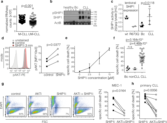
Targeted Pi3k Akt Hyperactivation Induces Cell Death In Chronic Lymphocytic Leukemia Nature Communications
Https Www Childrensmn Org References Lab Flowcyt Leukemia Lymphoma Panel Pdf
Immunophenotyping Utilizing 6 Color Flow Cytometry Warde Medical Laboratory

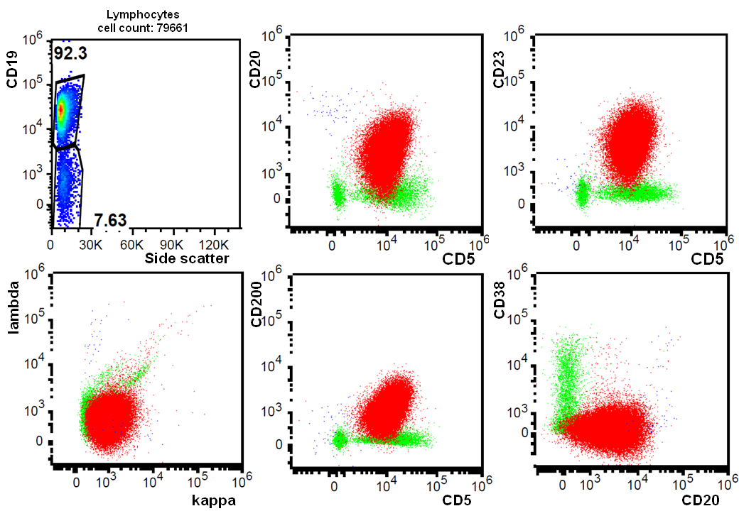
Chronic Lymphocytic Leukemia Small Cell Lymphoma Cll Sll Flow Cytometry
Acute T Lymphoblastic Leukemia Lymphoma T All Flow Cytometry

International Clinical Cytometry Society
Pin On Cd Markers
Pathology Outlines Cll Sll

Pin On Medical Laboratory Science

Flow Cytometry In The Diagnosis Of Mature B Cell Lymphoproliferative Disorders Debord 2020 International Journal Of Laboratory Hematology Wiley Online Library

Non Hodgkin Lymphoma Cancer Network Tabelle Diagramm Marker

Flowchart Diagram Of The Euroflow Strategy For Immunophenotypic Download Scientific Diagram

Pin By Cristin Lynn Perna On Laboratory Stuff Medical Laboratory Medical Laboratory Science Medical Technology

Reproducible Diagnosis Of Chronic Lymphocytic Leukemia By Flow Cytometry An European Research Initiative On Cll Eric European Society For Clinical Cell Analysis Escca Harmonisation Project Rawstron 2018 Cytometry
Introduction To Flow Cytometric Analysis Flow Cytometry
Features Of Immunophenotypic Finding B Cell Lymphoproliferative Diseases By Flow Cytometry Chuksina Kazan Medical Journal

B Cell Leukemia Lymphoma Panel

Uw Laboratory Medicine Hematology Division
Post a Comment for "Flow Cytometry Leukemia Lymphoma Panel"